Une radiographie du crâne est une image des os qui entourent le cerveau, y compris les os du visage, le nez et les sinus.
Radiographie du crâne ; Radiographie de la face ; Radiographie de la tête
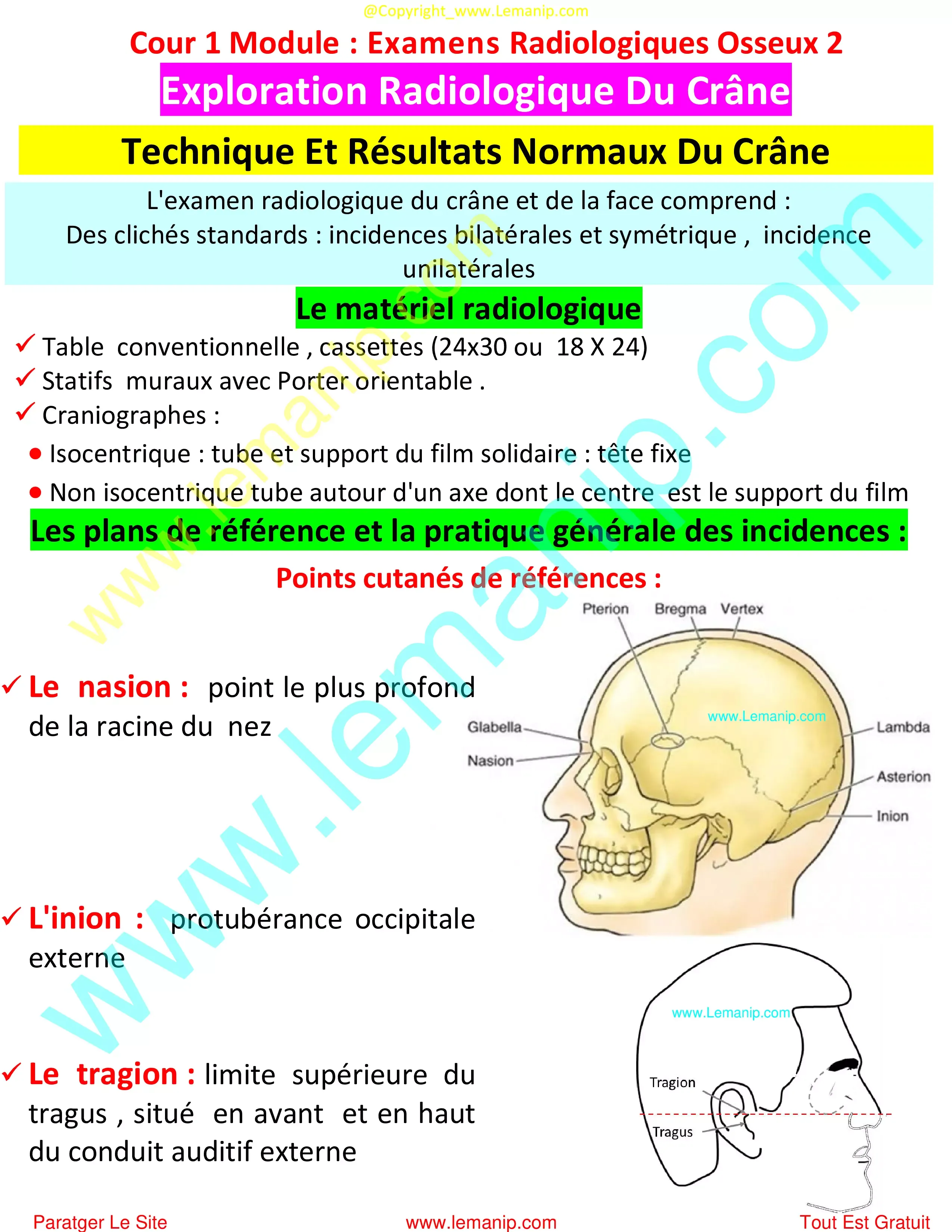
Exploration Radiologique Du Crâne
Cour 1 Incidences Radiologiques Du CrâneModule : Examens Radiologiques Osseux 2Manipulateur en Radiologie 2eme Année Semestre 3 Paramédical
1. Exploration Radiologique Du Crâne
2. Technique Et Résultats Normaux Du Crâne
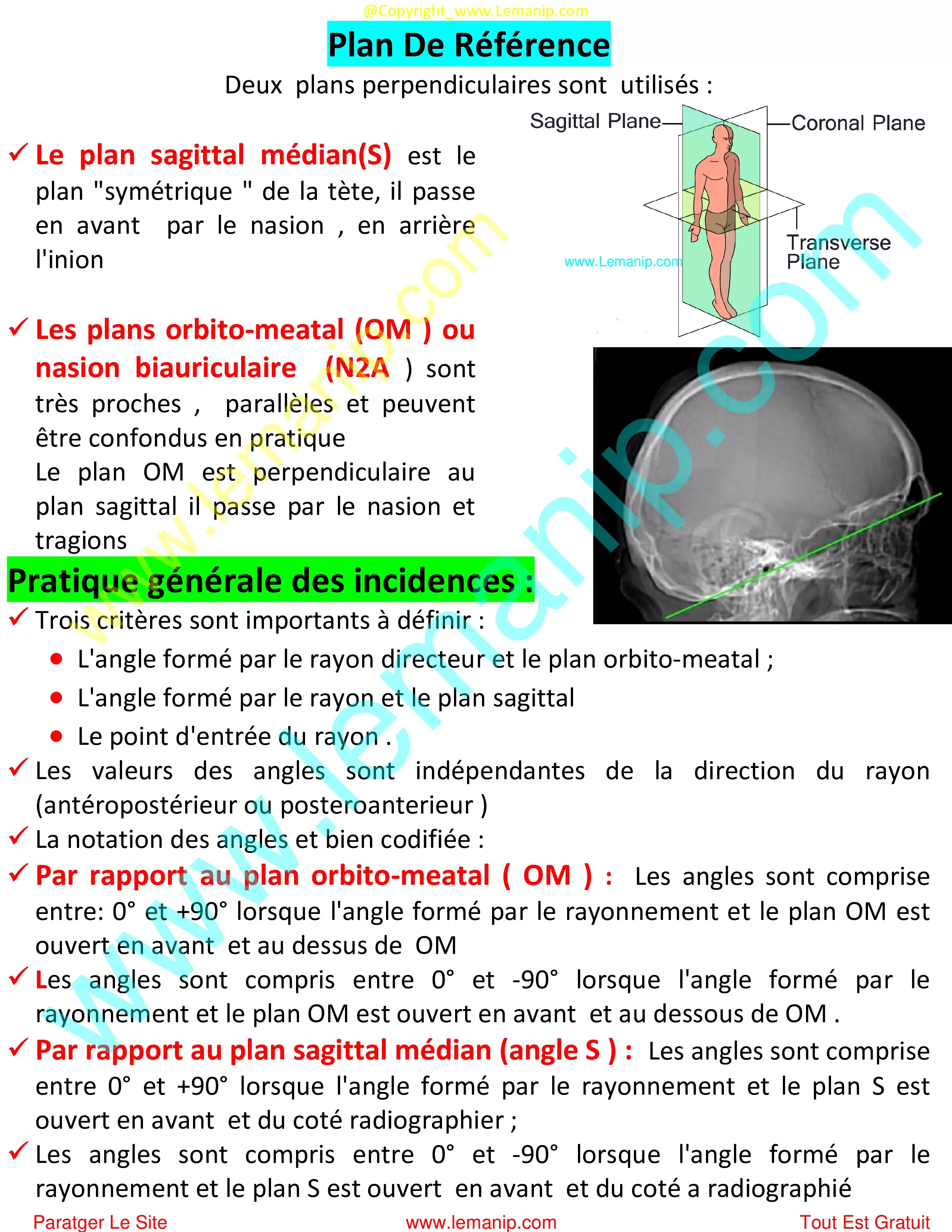
Plan De Référence
3. Radiographie Incidences Bilatérales Du Crâne
Cliquer Sur Next pour continuer la lecture et voire la page suivante
↚
A la fin il y a tout les autre cours et tous les modules
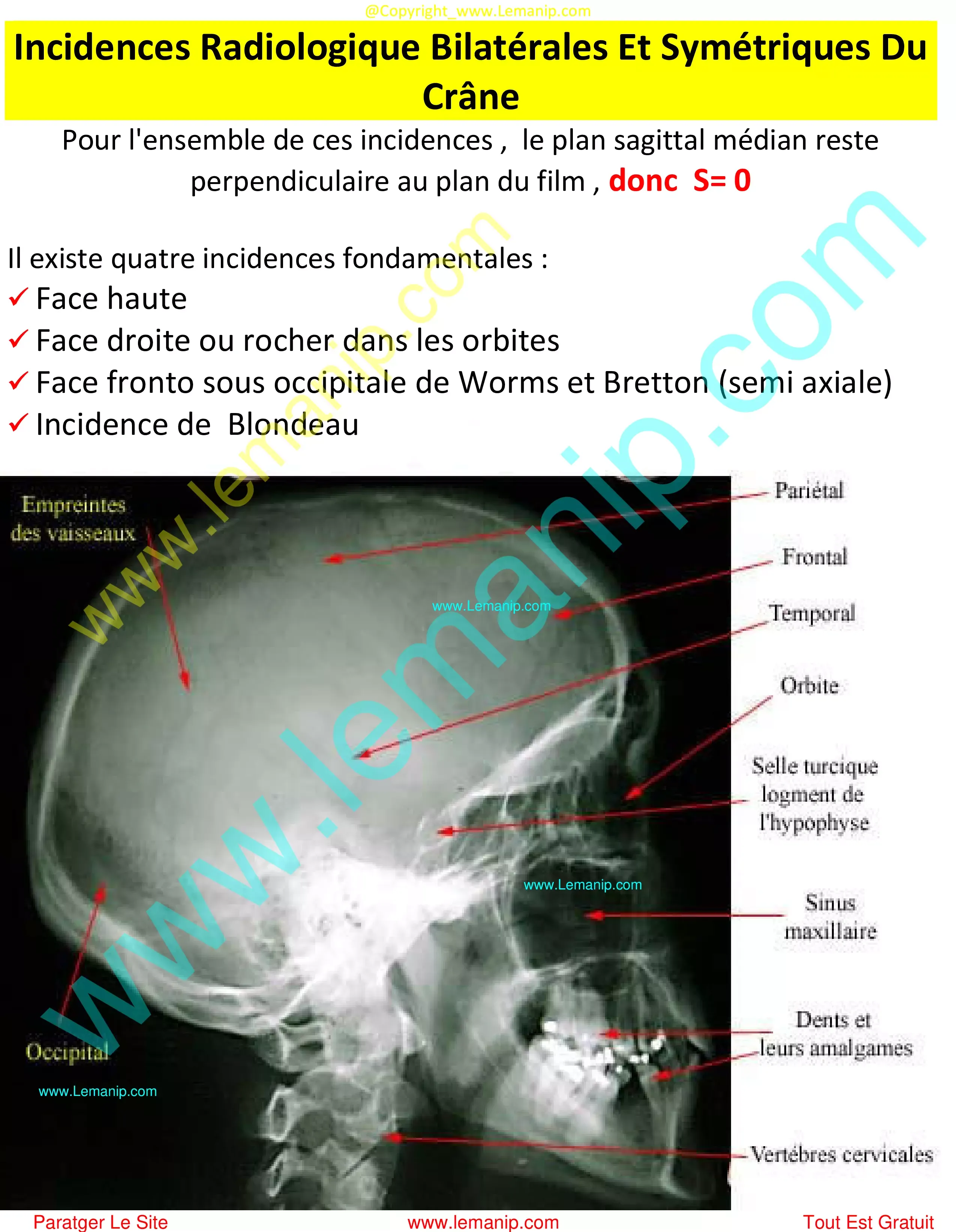
3. Incidences Radiologique Bilatérales Et Symétriques Du Crâne
incidence de face haute (Caldwell'S)
Face Droite Ou Rochers Dans Les Orbites
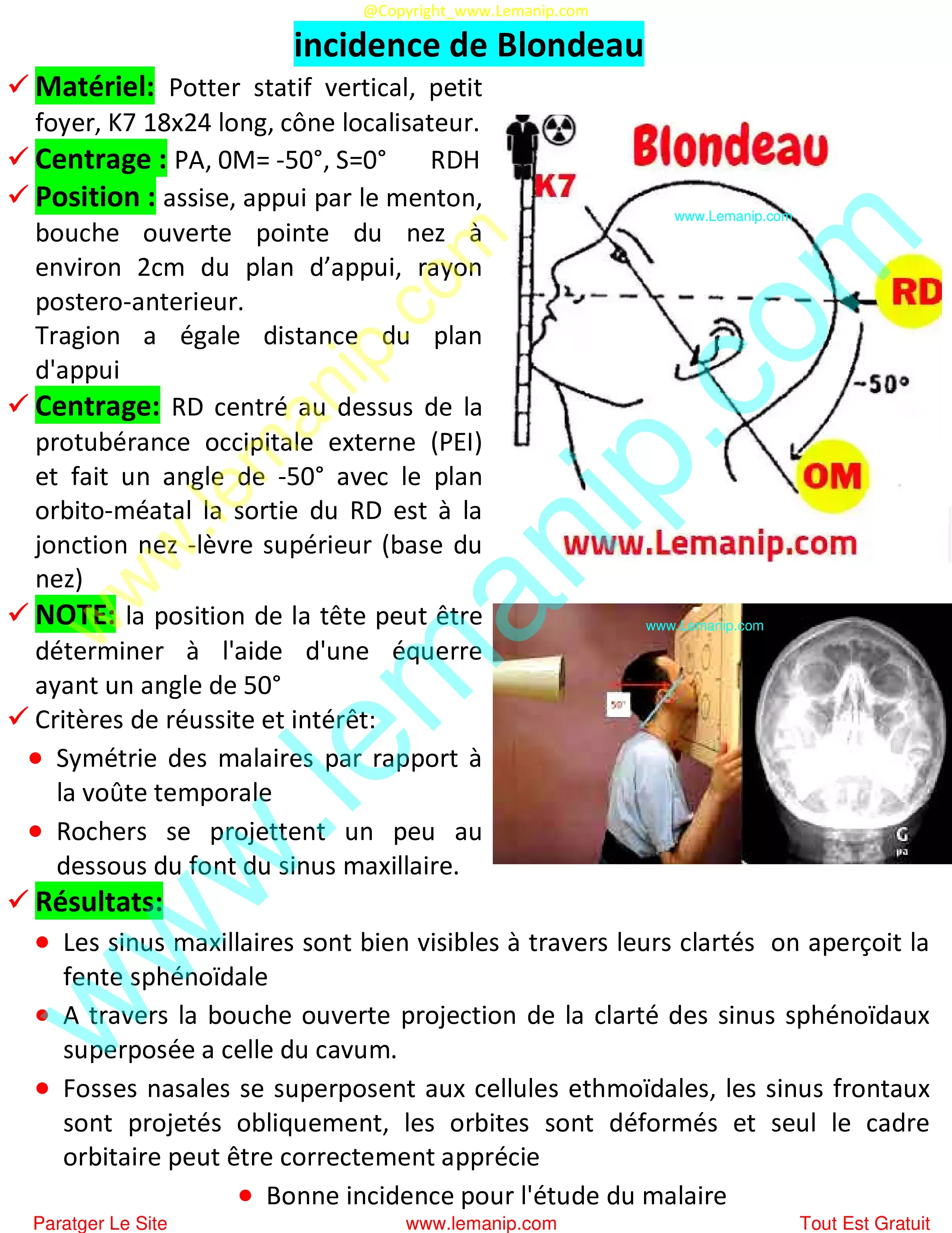
incidence de Blondeau
Incidence fronto sous occipital Worms et Bretton
incidence avec appui frontal : face basse
Les Trous Déchirés Postérieurs (TDP)
incidence axiales
4. Radiographie Incidences Unilatérales Du Crâne
Cliquer Sur Next pour continuer la lecture et voire la page suivante
↚
A la fin il y a tout les autre cours et tous les modules
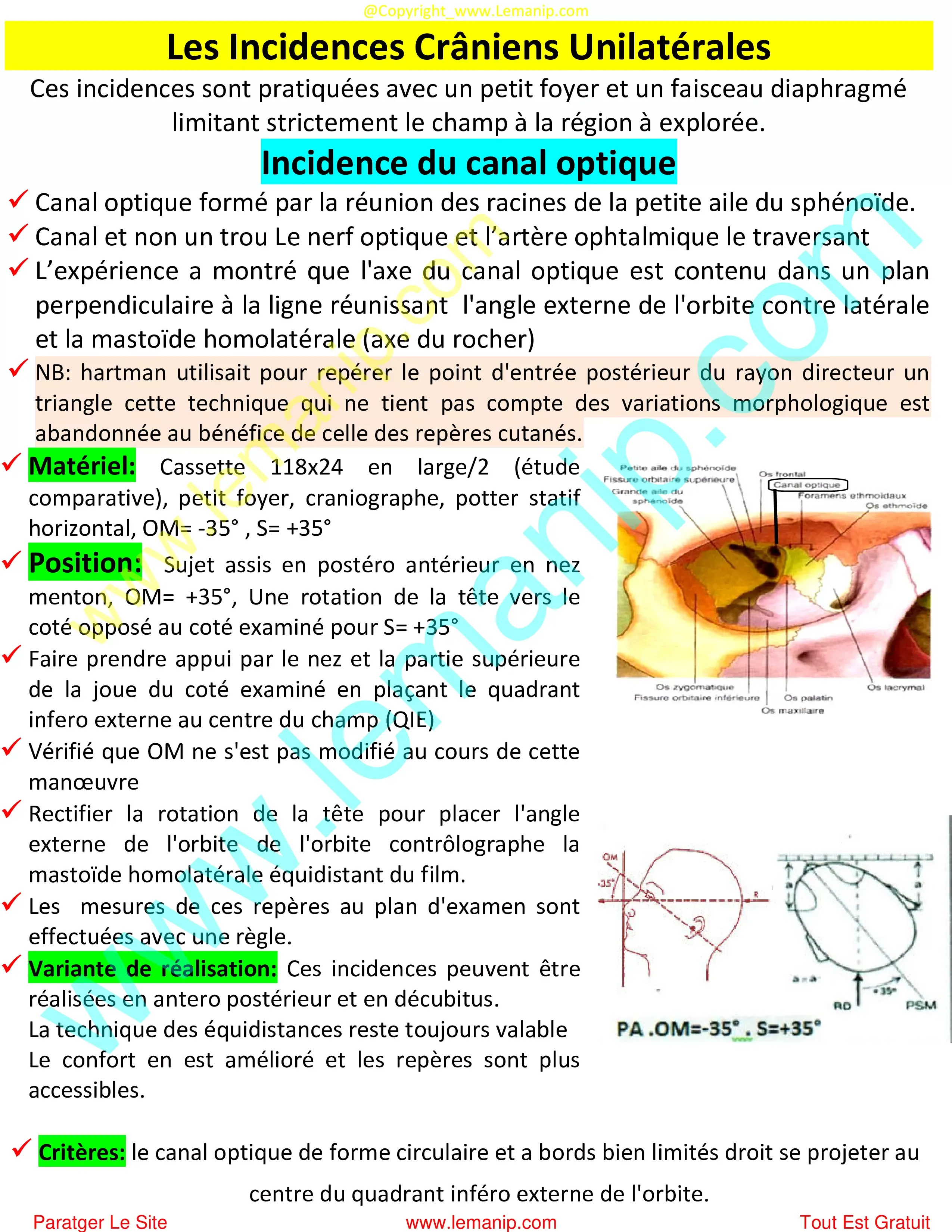
4. Les Incidences Crâniens Unilatérales
Incidence De La Fente Sphénoïdal : incidence De Brunetti
5. Radiographie Incidences Du Temporal
Cliquer Sur Next pour continuer la lecture et voire la page suivante
↚
A la fin il y a tout les autre cours et tous les modules
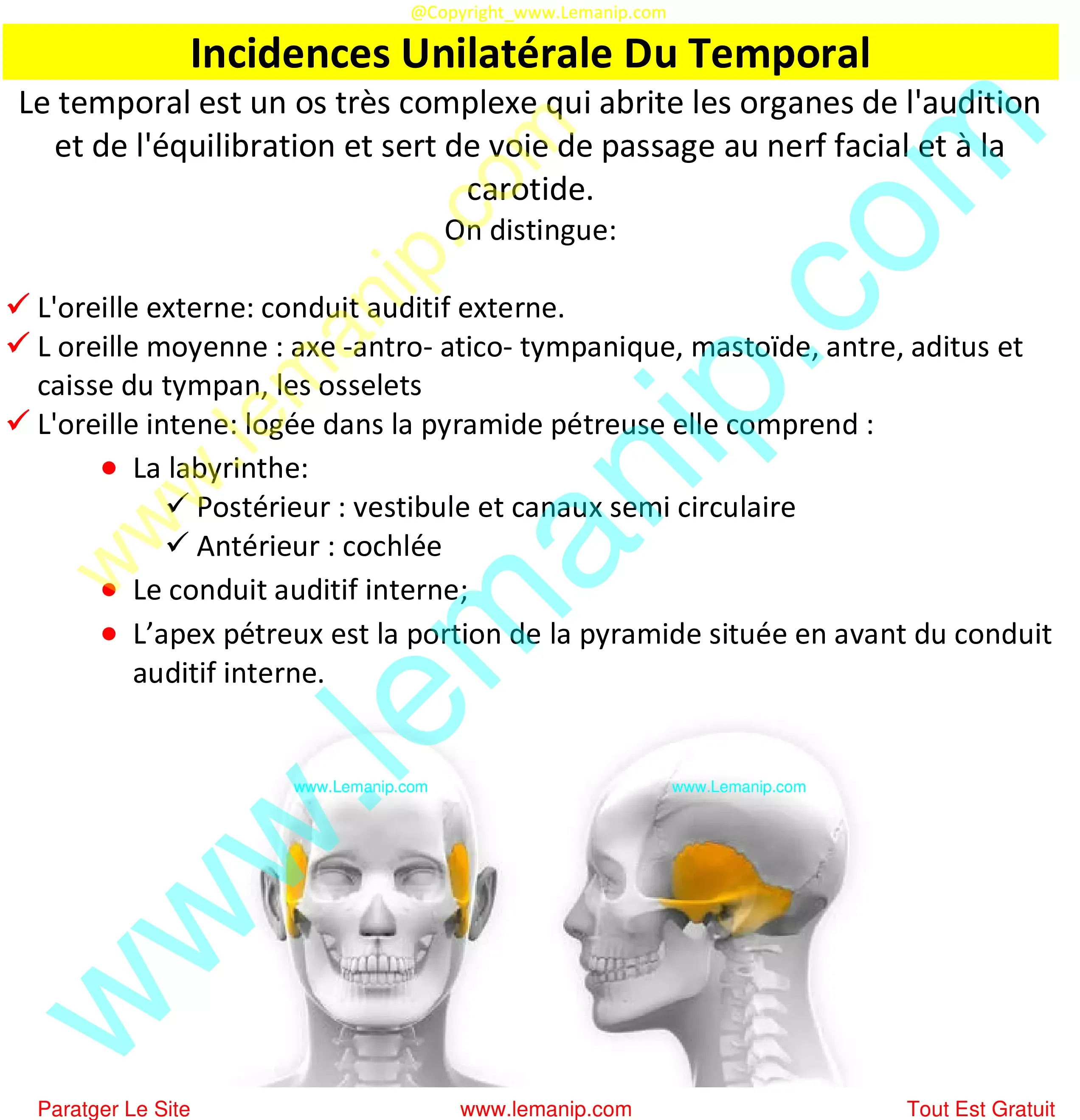
5. Incidences Unilatérale Du Temporal
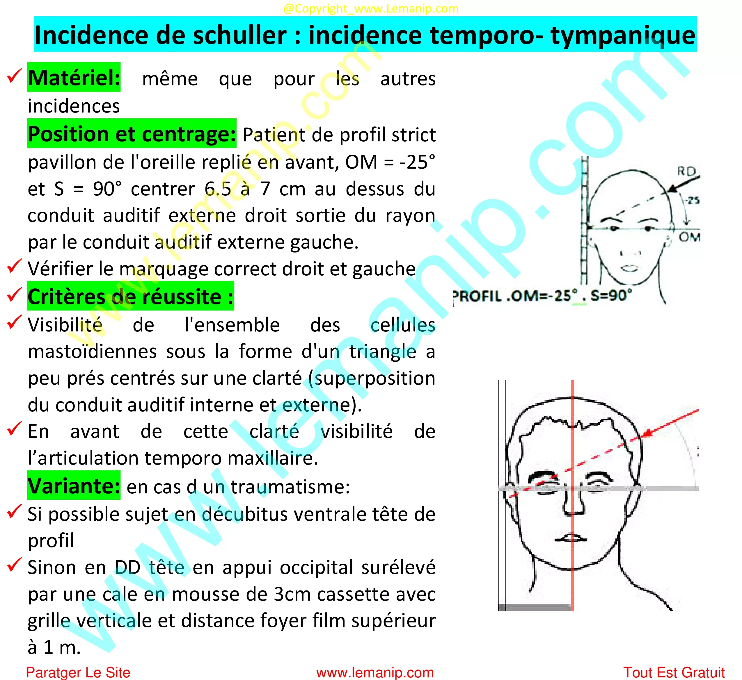
Incidence de schuller : incidence temporo- tympanique
Incidence de stenvers : occipito zygomatique
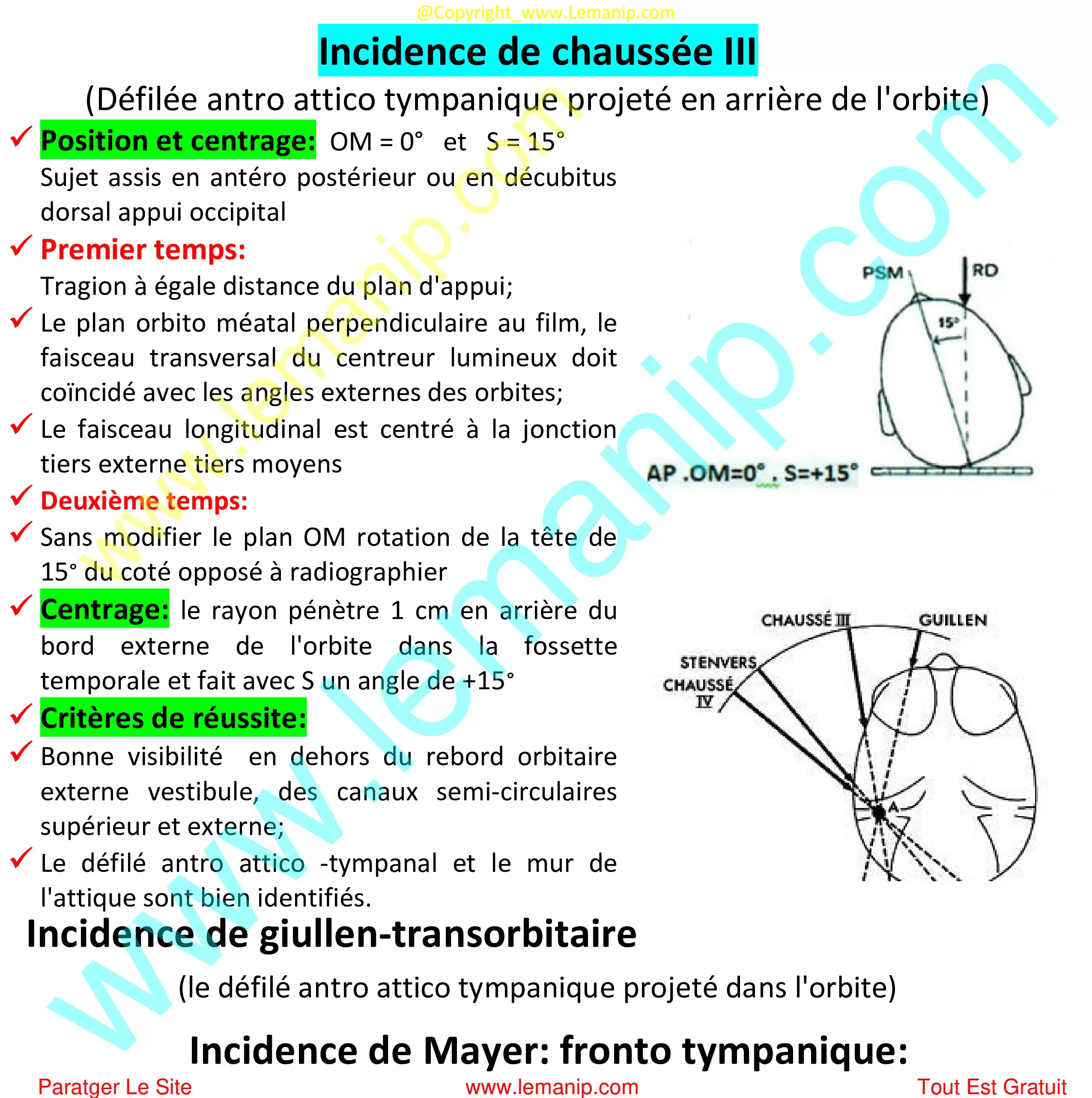
Incidence de chaussée III
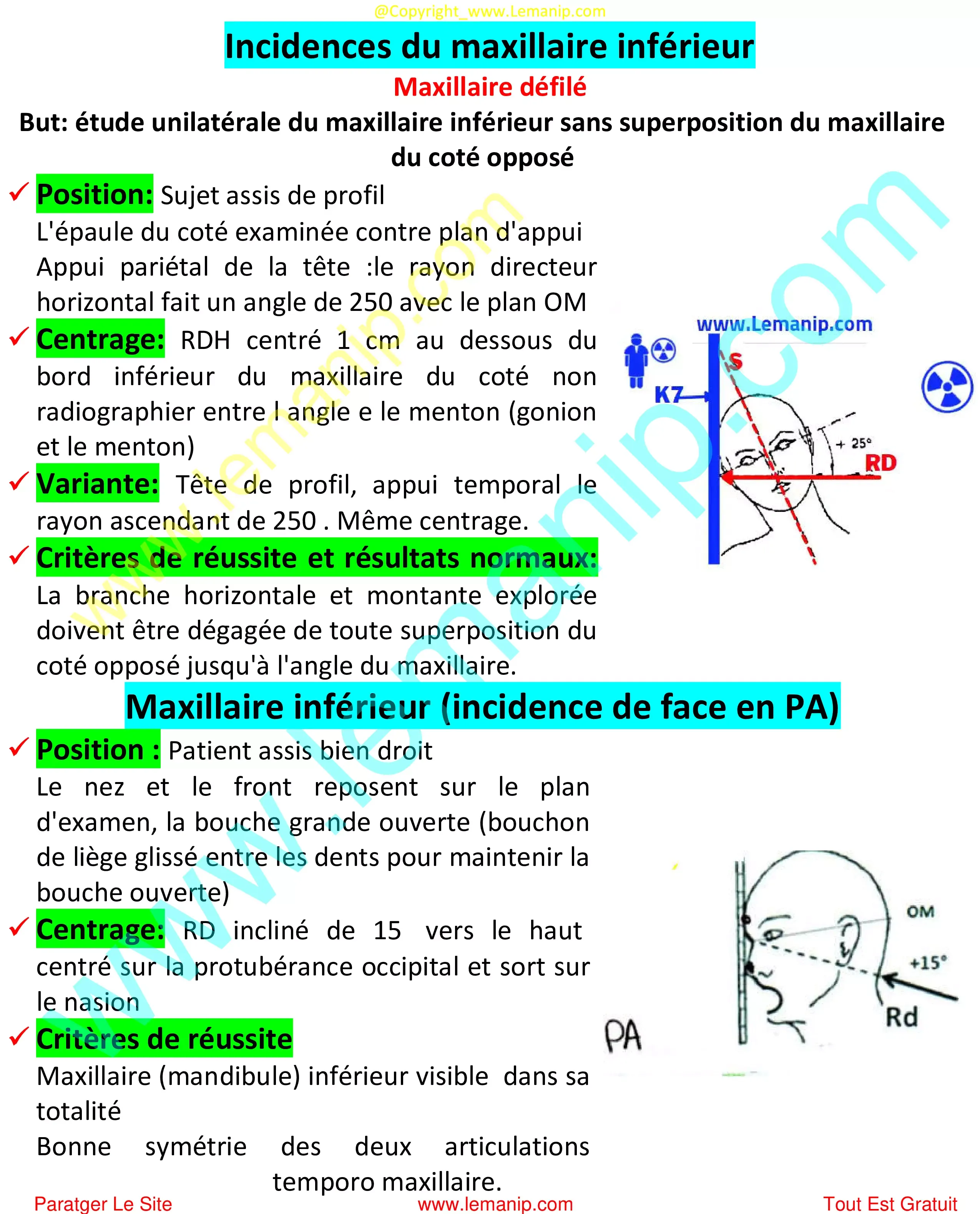
Incidences du maxillaire inférieur
Symphyse Mentonnière
Arcade zygomatique
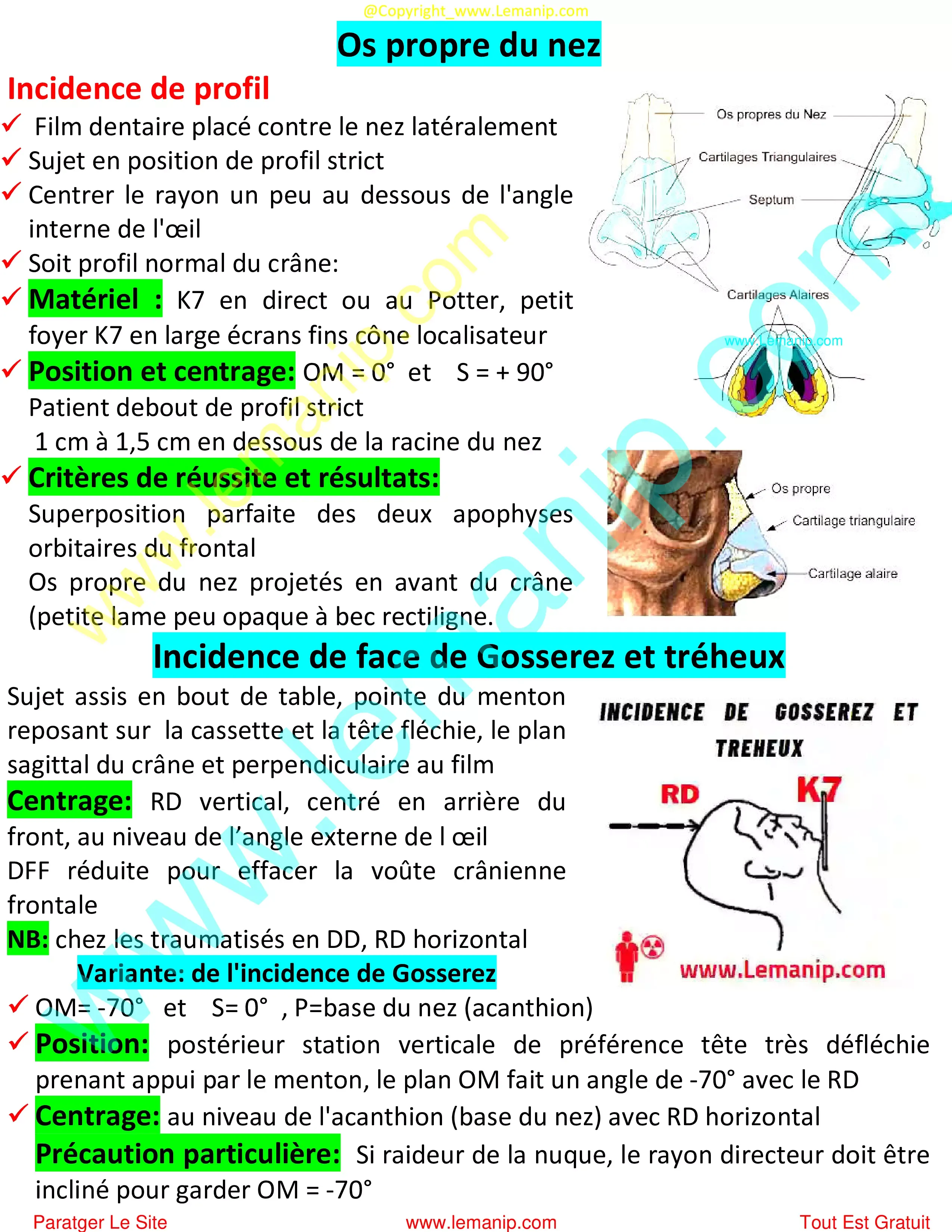
Os propre du nez
Articulation Temporo Maxillaire
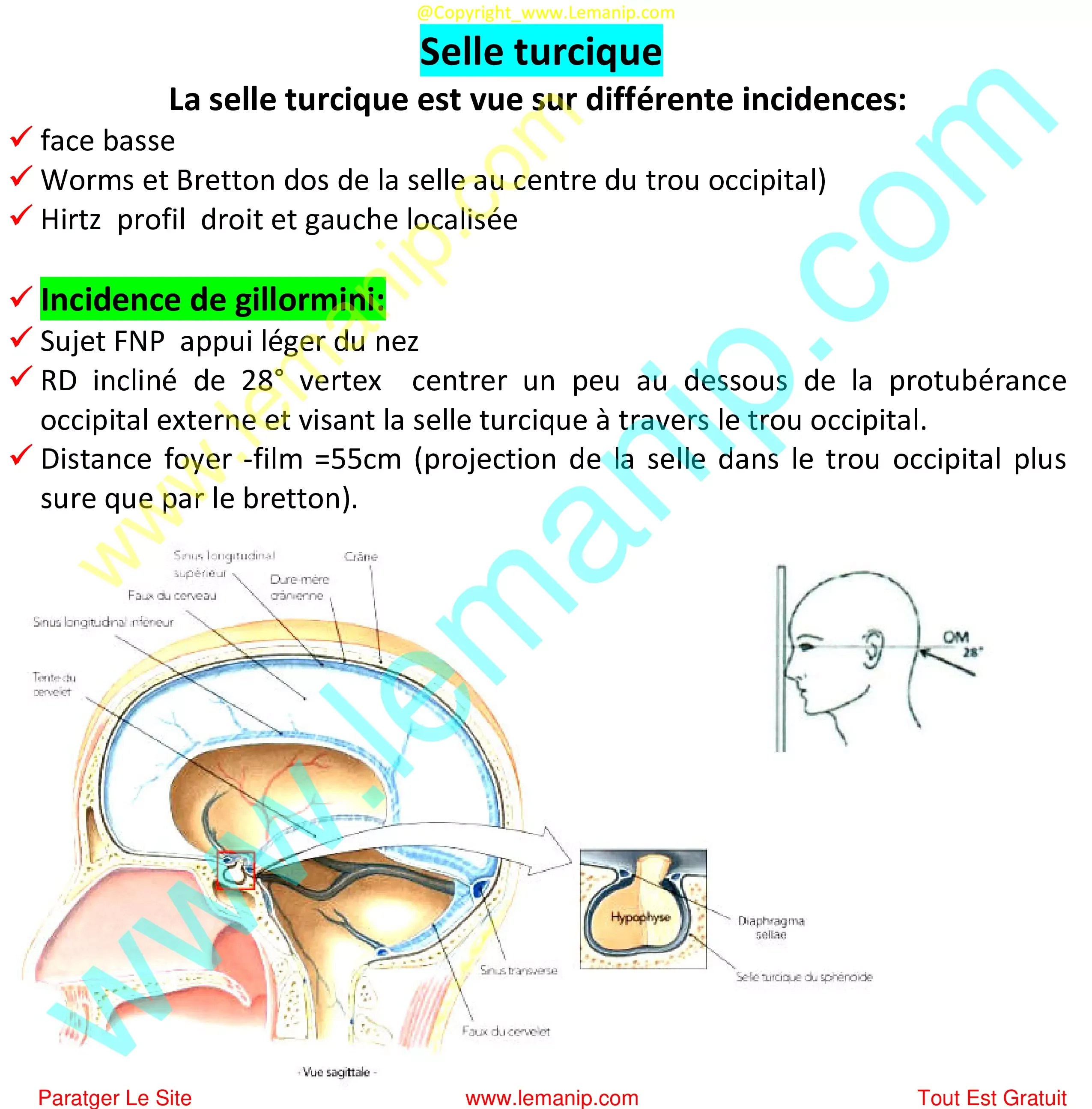
Selle turcique
Etude tangentielle de la voûte
Cliquer Sur Next pour continuer la lecture et voire la page suivante
↚
A la fin il y a tout les autre cours et tous les modules
6. Radiographie Panoramique
Intérêts, Indication Et Contre Indication De La Panoramique Dentaire
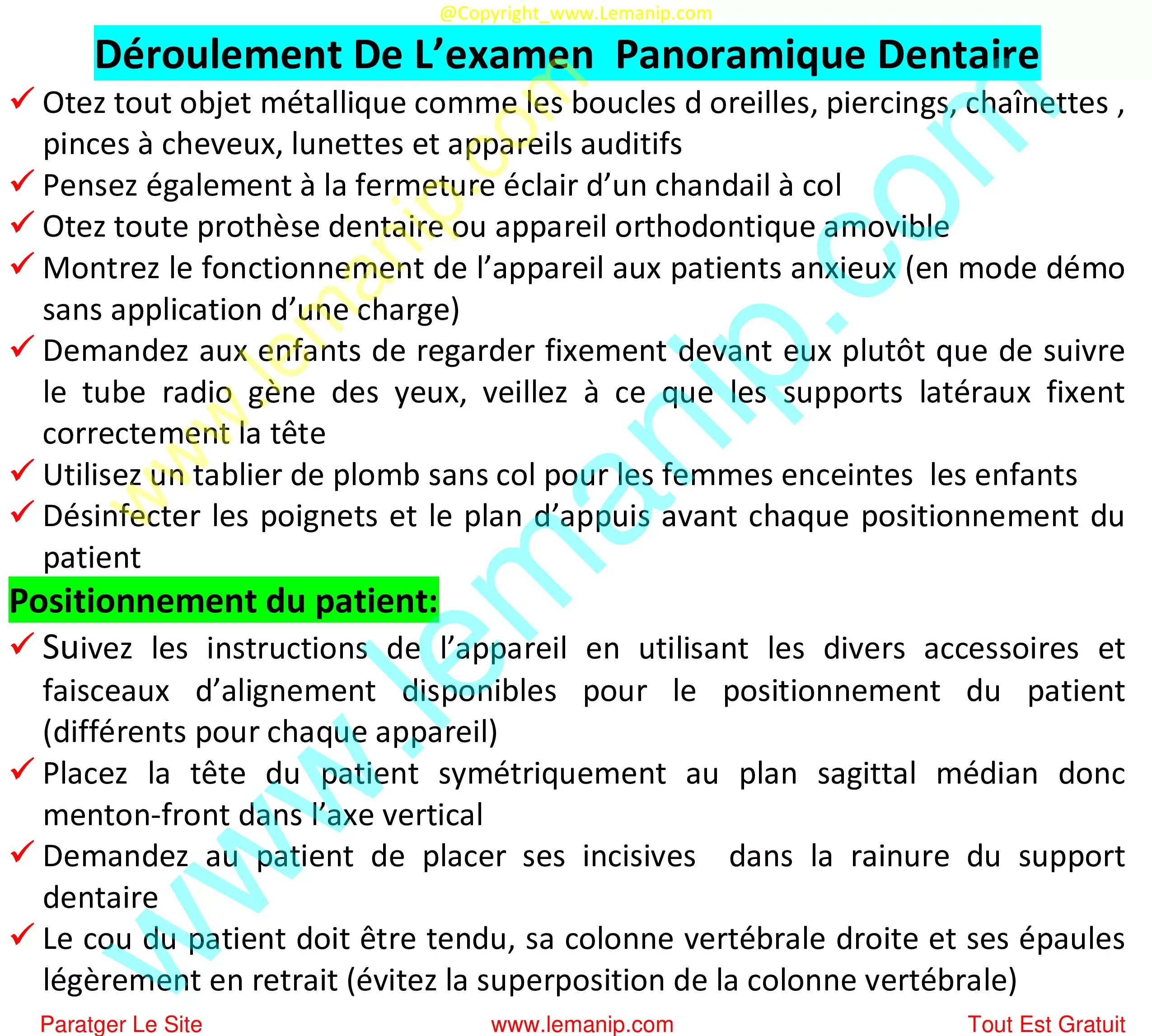
Déroulement De L’examen Panoramique Dentaire
7. Résumé Du Cour 1 Du Module Examens Radiologiques Osseux 2
Cours Du 3ème Année Manip en Radiologie
Module : IRM Physique Appliquée
- Cours ☢: ici Completement
- Cours ☢: Artéfacts En IRM Et Qualité d'image RM
- Cour 01: Introduction : La Radiologie Interventionnelle
- Cour 02: Angiographie Numérisée
- Cour 03: Angiographie Cérébrale
- Cour 04: La Coronarographie
- Cour 05: Artériographie Rénale
- Cour 06: Artériographie Des Membres Inférieurs
- Cour 07: Stéréotaxie Cérébrale
- Cour 08: Radiologie Interventionnelle En Oncologie
- Cour 09: Vertebroplastie En Cancérologie
- Cour 10: Chimio Embolisation
- Cour 11: Angioplastie Coronaire
- Cour 12: Ponction Ascitique
- Cour 01: Dosimetrie Explication Des Experts
- Cour 02: Dosimetrie : Physique Fondamentale
- Cour 03: Interaction Avec La Matière
- Cour 04: Dosimétrie De Photon De Haute Énergie
- Cour 05: Dosimetres et la Dosimetrie
- Cour 01: Rappel Du Système Nerveux
- Cour 02: Électromyographie EMG Et EMNG
- Cour 03: Électro-Encéphalographie EEG
- Cour 04: Les Potentiels Évoqués
- Cour 05: Électrocardiographie ECG
- Cour 01: Radiobiologie : Radiothérapie
- Cour 02: Radiothérapie Externe
- Cour 03: Curiethérapie: Radiothérapie
- Cour 04: Accessoires De Radiothérapie
- Cour 05: Système De Planification
- Cour 06: Immobilisation Et Contention
- Cour 07: Techniques D’irradiation Standards
- Cour 08: Techniques Innovantes En Radiothérapie
- Cour 09: Place De La Radiothérapie Dans Le Traitement
- Cour 10: Machines De Traitement
- Cour 11: Radiothérapie de contact 1
- Cour 12: Contrôle De Qualité
- Cour 13: Chaine De La Radiothérapie
- Cour 14: Radiothérapie de contact 2
- Cour 01: Réseaux Informatiques Radiologique
- Cour 02: Systèmes D’information Médicale Et Hospitalier
- Cour 03: Systèmes D’information RIS Et PACS
- Cour 04: DICOM (HL7, IHE, HIS, RIS, PACS)
- Cours ☢: ici Completement
- Cour 01: Contrôle Qualité En Imagerie Médicale
- Cour 02: Contrôle qualité en imagerie a modalité Rx
- Cour 03: Contrôle Qualité En Radiologie Conventionnelle
- Cour 04: Contrôle Qualité En Mammographie
- Cour 05: Contrôle Qualité En Scannographie
- Cour 06: Contrôle Qualité En Radiothérapie 1
- Cour 07: Contrôle De Qualité En Radiothérapie 2
- Cour 01: Introduction Au Médecine Nucléaire
- Cour 02: Principes Du Médecine Nucléaire
- Cour 03: TEP Scan (TEP-CT)
----------------------------------------------------------------------
Ne jamais oublier que
Un examen radiologique correct
(manipulation, position, condition et critère de réussite)
- Implique des cliches et images radiographiques de bonne qualité facile à interpréter
- Implique une interprétation correcte et un vrai diagnostique à poser
- Donc un traitement efficace qui doit arrêter les douleurs et les souffrances des patients
Donc soyez responsable c’est le temps de devenir un pro dans la radiologie manipulation et interprétation
Suivez-nous et partagez tout est gratuit, il y a plusieurs collègues qui sont besoins de nous
Merci à vous
----------------------------------------------------------------------
- Cour 01: Exploration Radiologique Des Poumons
- Cour 02: Sialographie : Exploration Rx Glandes Salivaires
- Cour 03: Abdomen Sans Préparation ASP
- Cour 04: Exploration Rx Digestif :TOGD TGD LB TG TO
- Cour 05: Exploration Rx Urinaire : UIV UCR AUSP
- Cour 06: Cholangiographie : Exploration Rx Voies Biliaires
- Cour 07: Bronchographie Opaque
- Cour 08: Exploration Radiologique Du Larynx
- Cour 01: La Nutrition
- Cour 02: Adaptation Des Besoins Nutritionnels
- Cour 03: Equilibre Alimentaire
- Cour 04: Groupes Des Aliments
- Cour 05: Etudes Des Différents Groupes Des Aliments
- Cour 06: Principales Maladies Carentielles
- Cour 01: Groupes Sanguins Et Système Rhésus
- Cour 02: Hémostase
- Cour 03: FNS : Hémogramme
- Cour 04: Fibrinogène
- Cour 05: Bilan Rénal
- Cour 06: ECBU : Examen Cyto Bactériologique Urinaires
- Cour 07: Ionogramme Sanguin
- Cour 08: Glycémie
- Cour 09: Gaz Du Sang
- Cour 10: Bilan Lipidique
- Cour 11: La Bilirubine
- Cour 12: Bilans Biologiques : Indications Et Normes
- Cour 01: Techniques Radiologiques Pulmonaires
- Cour 02: Explorations Radiologiques De L'appareil Urinaire
- Cour 03: Etude Statiques Du Membre Inférieure
- Cour 04: Luxation Congénitale De La Hanche
- Cour 01: Sémiologie Pathologie De L'appareil Cardiovasculaire
- Cour 02: Sémiologie Pathologie Du Système Nerveux
- Cour 03: Sémiologie Pathologie Génito-Urinaire
- Cour 04: Sémiologie Pathologie Endocrinienne
- Cour 05: Sémiologie Pathologie Mammaire
- Cour 06: Sémiologie Pathologie En ORL
- Cour 01: Introduction A La Pharmacologie Radiologique
- Cour 02: Produit De Contraste Baryté
- Cour 03: Produits de Contraste Iodés PCI
- Cour 04: Produit De Contraste En Échographie
- Cour 05: Produit De Contraste En IRM
- Cour 06: Les Antispasmodiques
- Cour 07: Les Antalgiques
- Cour 08: Les Antiseptiques
- Cour 09: Les Antibiotiques
- Cour 10: Les Anti Inflamatoires
- Cour 11: Les Anti Coagulant
- Cour 12: Allergie, État De Choc Et Collapsus
- Cour 13: Choc Anaphylactique
- Cour 01: Environnement De Travail Informatique
- Cour 02: Système D'exploitation Windows
- Cour 03: Microsoft Office Word
- Cour 04: Microsoft Office Excel
- Cour 05: Microsoft Office PowerPoint
- Cour 01: Mammographie : Exploration Rx Des Seins
- Cour 02: Galactographie
- Cour 03: Biopsie Mammaire Stéréotaxique
- Cour 04: Fistuloographie
- Cour 05: Hystérosalpingographie : HSG
- Cour 06: Arthrographie
- Cour 07: Déférentographie : Stérilité Chez L’homme
- Cours ☢: ici Completement
- Cour 01: Mécanisme De La Cancérogénèse
- Cour 02: Introduction A La Cancérologie
- Cour 03: Évolution Du Cancer
- Cour 04: Prévention Et Dépistage Des Cancers
- Cour 05: Diagnostic Du Cancer
- Cour 06: Classification Du Cancer
- Cour 07: Traitements Du Cancer
- Cour 08: Prise En Charge Psychologique Des Cancéreux
- Cour 01: Description Du métier Manip en Radiologie
- Cour 02: Manipulateur en Scanner TDM
- Cour 03: Fondements de la profession Manip en Radiologie
- Cour 00: La Cellule
- Cour 01: Etude Morphologique De La Cellule
- Cour 02: Matériel Génétique
- Cour 03: Etapes de la division cellulaire
- Cour 04: Aberration Chromosomique
- Cour 05: Hérédité et mode de transmission des maladies
- Cour 06: Notions D'Embryologie
- Cour 07: Ostéologie
- Cour 08: Les Membres Supérieurs
- Cour 09: Le Rachis
- Cour 10: Les Membres Inférieurs
- Cour 11: Le Crâne
- Cour 12: Appareil Locomoteur
- Cour 13: Arthrologie
- Cour 14: La Myologie
- Cour 15: Le Squelette
- Cour 16: Appareil Respiratoire
- Cour 17: Histologie De L'appareil Respiratoire
- Cour 18: Anatomie De L’appareil Digestif
- Cour 19: Physiologie De L’appareil Digestif
- Cour ☢: ici Completement
- Cour 01: Definition Du Secourisme
- Cour 02: Dégagement d'urgence et Surveillance
- Cour 03: Arrêt Cardio Respiratoire (Detress respiratoire)
- Cour 04: Le Malaise
- Cour 05: Brûlure Secourisme
- Cour 06: Plaies-Simple et Grave
- Cour 07: Traumatismes-Secourisme Des Os
- Cour 08: Hémorragie
- Cour 09: Bandages
- Cour 10: Brancardage
- Cours ☢: Techniques de soins
- Cour 01: Techniques d’expression écrite et orale
- Cour 02: Comment rédiger une bibliographie ?
- Cour 03: Etapes Recherche Documentaire Efficace
- Cour 04: Comment faire un exposé oral ?
- Cour 05: Types de phrases
- Cour 06: Pronom Interrogatif
- Cour 07: Pronom Possessif
- Cour 08: Pronom Relatif
- Cour 09: Connecteur /Articulateurs Logiques
- Cour 01: Definition de la pharmacie
- Cour 02: Pharmacologie Générale
- Cour 03: Formes pharmaceutiques et voies d'administration
- Cour 04: Facteurs influencant la reponse d'un medicament
- Cour 05: Conseils d'utilisation des médicaments et DMs
- Cour 06: Pharmacie hospitalière
- Cour 07: Activité, Toxicité Et Interactions Des Medicaments
- Cour 01: Introduction à la Santé Publique
- Cour 02: Introduction à l'épidémiologie
- Cour 03: Branches de l'épidémiologie et indicateurs
- Cour 04: Mesures d'associations
- Cour 05: Sécurité Sociale
- Cour 06: Financement de la santé
- Cour 07: Système de santé dans le monde
- Cour 08: Systéme National De Santé
- Cour 09: Liste des maladies à déclaration obligatoire + exo SP
- Cour ☢: ici Completement
- Cour ☢: Loi Du RadioProtection en Algerie COMENA
- Cour ☢: Loi des manipulateurs et santé En Algerie
- Cour 01: Introduction A La Psychologie
- Cour 02: La Personnalité
- Cour 03: Mécanismes De Défense Et D'adaptation
- Cour 04: Relation Soignant Soigné
- Cour 05: États Affectif
- Cour 06: La Communication
- Cour 07: Trarnsfert Et Le Contre Trarnsfert
- Cour 01: Microbiologie - Le Monde Microbien
- Cour 02: Epdemiologie Des Maladies Transmissibles
- Cour 03: Infection
- Cour 04: Infections Nosocomiales - Epidemiologie
- Cour 05: Immunité : Explication
- Cour 06: Isolement : Explication
- Cour 07: Concept D’hygiène
- Cour 08: Prevention : Explication
- Cour 09: Déchets hospitaliers : Types et risques pour la santé
- Cour 01: Rayons X : Nature, Domaine Et Découverte
- Cour 02: Propagation Et Absorption Des Rayons X
- Cour 03: Production Et Emission Des Rayons X
- Cour 04: Générateur Rayons X
- Cour 05: Pupitre De Commande : Rayons X
- Cour 06: Tube à Rayons X
- Cour 07: Vieillissement et accidents du tube a rayon X
- Cour 08: Gaine Et Annexes Au Tube A Rayons x
- Cour 09: Statifs D'utilisation
- Cour 10: Grille Anti Diffusante
- Cour 11: Injecteurs Automatiques Scanner et IRM
- Cour 12: Amplificateur De Brillance ( Luminance )
- Cour 13: Détecteurs des images radiologiques
- Cour 01: Notions De Base De L'électricité
- Cour 02: Rayonnement Electromagnétique
- Cour 03: Structure De La Matiere et L'atome
- Cour 04: La Radioactivité
- Cour 05: Interactions (RI) Avec La Matière
- Cour 06: Interaction Rayonnement Électromagnétique-Matière
- Cour 07: Détection Des Rayonnements Ionisants
- Cour 08: Dosimetrie : Physique Fondamentale
- Cour 01: Formation Géométrique De L’image Radiologique
- Cour 02: Contraste En Radiologie
- Cour 03: Flou Et Netteté Radiologique
- Cour 04: Materiel Radiographique Radiologique
- Cour 05: Traitement Chimique Du Film Radiographique
- Cour 06: Constantes Et Paramètres D’exposition Radiologique
- Cour 07: Méthodes Radiologiques
- Cour 08: Numérisation : Imagerie Digitale
- Cour 01: Généralités Sur Les Incidences Radiologiques
- Cour 02: Incidences Radiologiques De L'épaule, Omoplate et Clavicule
- Cour 03: Incidences Radiologiques De L'Humérus, Coude et La Main
- Cour 04: Incidences Radiologiques Du Bassin et L'Hanche
- Cour 05: Incidences Radiologiques Du Femur Et Genou
- Cour 06: Incidences Radiologiques Du Jambe,Cheville Et Pied


