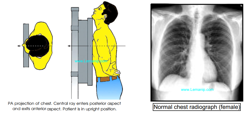
Chest x-ray PA View, Posterior thoracic examination (PA) examines the lungs, the bony part of the thoracic cavity, the mediastinum, and the main vessels.

Indications of Chest PA
Chest X-ray is the most common X-ray examination in the emergency department. PA Radiography is often used to help diagnose a number of acute and chronic diseases affecting all organs of the thoracic cavity.
In addition, it serves as the most sensitive simple radiograph for detecting free intraperitoneal gas or pneumoperitoneum in patients with acute abdominal pain.
Patient position in Chest xray PA View
✔️ The is erect facing the upright image receptor, the superior aspect of the receptor is 5 cm above the shoulder joints
✔️ The chin is raised as to be out of the image field
✔️ The shoulders are rotated anteriorly to allow the scapulae to move laterally off the lung fields, and this can be achieved by either:
- Hands placed on the posterior aspect of the hips, elbows partially flexed rolling anterior or
- Hands are placed around the image receptor in a hugging motion with a focus on the lateral movement of the scapulae
✔️ The shoulders are depressed to move the clavicles below the lung apices
Technical factors chest xray pa
Posteroanterior projection
✔️ PA
Suspended inspiration
Centering point
✔️ The level of the 7th thoracic vertebra, approximately the inferior angle of the scapulae
collimation
✔️ superiorly 5 cm above the shoulder joint to allow proper visualization of the upper airways
✔️ inferior to the inferior border of the 12th rib
✔️ lateral to the level of the acromioclavicular joints
Orientation
✔️ portrait or landscape
Detector size
✔️ 35 cm x 43 cm or 43 cm x 35 cm
Exposure
✔️ 100-110 kVp
✔️ 4-8 mAs
SID
✔️ 180 cm
Grid
✔️ yes
Technical evaluation of the image Chest PA
✔️ All pulmonary fields should be visible from the apices to the lateral costo-occipital corners.
✔️ The entire lung fields should be visible from the apices down to the lateral costophrenic angles.
✔️ The chin should not be superimposing any structures
✔️ Arms are not superimposed over the lateral chest wall (this can mimic pleural thickening)
✔️ Minimal to no superimposition of the scapulae borders on the lung fields
✔️ Sternoclavicular joints are equidistant from the spinous process
✔️ The clavicle is in the same horizontal plane
✔️ A maximum of ten posterior ribs are visualized above the diaphragm
✔️ The 5th-7th anterior ribs should intersect the diaphragm at midclavicular line
✔️ The ribs and thoracic cage are seen only faintly over the heart
✔️ Clear vascular markings of the lungs should be visible

Practical points of cxr pa
The phase of respiration or breathing phase profoundly affects the appearance of several structures on the chest X-ray (images of inhalation and exhalation in the same patient are shown in Example 2). An X-ray of a PA with a bad breath can simulate pathology. Structures that may differ upon expiration include:
✔️ the size of the heart
✔️ the contours of the mediastinum and the width
✔️ Lung Inflation
✔️ the contours of the diaphragm and width
The rotation of the chest X-ray can mimic standard pathological processes and make it difficult to make an appropriate diagnosis.
The Chest PA view mode is used to study a variety of conditions, and the responsibility of the radiologist is to ensure stable receipt of high-quality diagnostic images.
The sternoclavicular joints are an audible indicator of positional rotation, if one sternoclavicular joint is noticeably wider than the other, this corresponding side must be rotated towards the image receptor in order to correct the rotation.
Patients with a history of prolonged emphysema or COPD will have abnormally long lungs compared to the general population, keep this in mind when comparing the upper and lower divisions.
The placement of side markers is mandatory; patients may have congenital diseases that mimic a mirror image2.
Remember to explain to your patient what you are going to do; that is, ask him to take a breath and hold it. In many cases, this gives the patient time to prepare and leads to better breath retention and, consequently, to a better X-ray image.
How to read a chest x ray
Always remember to tell your patient to breathe again! and
Chest x ray basic Interpretation

Chest x ray pa view Interpretation

Chest X-Ray
Chest X ray